ALTERED BONE MATRIX PROTEINS
DISORDERS IN COLLAGEN METABOLISM
Osteogenesis imperfecta (OI), also known as brittle bone disease, is a genetically determined bone disorder in which either defective or insufficient quantities of collagen molecules are produced. OI includes a group of clinically and genetically heterogeneous disorders characterized by high risk of bone fractures, with variable degree of severity and presumed or proven defects in collagen type 1 biosynthesis.
(OMIM phenotype number #166200)
Osteogenesis imperfecta (OI), also known as brittle bone disease, is a genetically determined bone disorder in which either defective or insufficient quantities of collagen molecules are produced. OI includes a group of clinically and genetically heterogeneous disorders characterized by high risk of bone fractures, with variable degree of severity and presumed or proven defects in collagen type 1 biosynthesis. Common clinical signs are: increased bone fractures, and secondary features such as short stature, blue sclerae, dentinogenesis imperfecta and hearing loss may also exist in affected individuals. The incidence of the different types of OI is approximately 1/15.000-20.000 births and most cases are due to autosomal dominant inheritance with mutations in collagen, type 1 alpha-1, type 1 alpha-2 (COL1A1 or COL1A2) genes, which encode the alpha 1 and alpha 2 chains of type 1 collagen. Mutations in COL1A1 and COL1A2 genes altering the structure or the amount of type 1 collagen, result in a skeletal phenotype that ranges from subclinical to lethal. Furthermore, there are several mutant noncollagen genes causing the 5–10% of recessive cases, such as CRTAP, LEPRE1, PPIB, PLOD2, FKBP10, SERPIN H1, SERPIN F1, BMP1, and IFITM5 genes. All types of OI cause fragile bone, which can include overmineralization or under-mineralization defects as well as abnormal collagen post-translational modifications.
The therapeutic management of OI needs various medical specialists (endocrinology, orthopedics, physiotherapy..). Medical therapies recommended include: bisphosphonates, potent antiresorptive drugs, for severe forms of OI, and supplementations of vitamin D and calcium. In case of bone and spinal deformities, and long bone fractures can be necessary surgical management. At last, it is recommendable physiotherapy in order to improve mobility and reduce the risk of fractures.
Gene
COL1A1 gene, 17q21.33 (OMIM gene/locus number +120150).
COL1A2 gene, 7q21.3 (OMIM gene/locus number *120160).
Phenotype
Mildest form of OI, due to 50 % reduction of the amount of collagen type I; blue sclerae, joint hyperextensibility, normal tooth (OI type IA, with a reduction in the amount of normal collagen) or dentinogenesis imperfecta (OI type IB, with abnormal collagen), hearing loss (onset usually around 20 years), mitral valve prolapse, thin skin, increased fracture rate throughout childhood (ensues when child begins to walk, decreases after puberty, then increases after menopause and in men aged 60–80 years), and biconcave flattened vertebrae.
Main biochemical alterations
No specific alterations in bone markers (low sclerostin, low PINP and PICP have been reported).
Other resources:
Perinatally lethal Osteogenesis Imperfecta (OI type II-A and II-B) is the most severe form of OI (See also OI type I). It is a lethal form with collagen abnormalities resulting in dwarfism, bone fragility, low bone mass, high risk of fractures (rib and long bone), and deformity with in utero or perinatal death.
(OMIM phenotype number #166210)
Perinatally lethal Osteogenesis Imperfecta (OI type II-A and II-B) is the most severe form of OI (See also OI type I). It is a lethal form with collagen abnormalities resulting in dwarfism, bone fragility, low bone mass, high risk of fractures (rib and long bone), and deformity with in utero or perinatal death. Some infants with type II OI may live for as long as a year, but eventually do succumb to multiple pneumonias or respiratory insufficiency. Early prenatal sonographic diagnosis is possible, because OI type II is a severe form with early fetal skeletal defects.
Type II-A
Gene: COL1A1 gene, 17q21.33 (OMIM gene/locus number +120150).
Gene: COL1A2 gene, 7q21.3 (OMIM gene/locus number *120160).
Type II-B
Gene: CRTAP gene, 3p22.3 (OMIM gene/locus number *605497).
Gene: LEPRE1 gene, 1p34.2 (OMIM gene/locus number *610339).
Gene: PPIB gene, 15q22.31 (OMIM gene/locus number *123841).
Phenotype
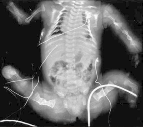
Most severe form of OI, neonatal lethality, born prematurely and small for gestational age, multiple neonatal fractures, shortening and bowing of long bones with severe under modeling leading to crumpled long bones, all vertebrae hypoplastic/crushed, hip abducted and knees flexed, severe osteoporosis with intrauterine fractures and abnormal modeling, skull with severe undermineralization with wide-open anterior and posterior fontanels, white or blue sclerae, death for respiratory insufficiency and pneumonias.
Fig. Radiograph of an infant with OI type II. It shows severe osteoporosis of skeleton with fractures of upper extremities, crumpled femora, flared rib cage with narrow apex and multiple beads of callus on each rib.
Reproduced from Marini J, Smith SM. Osteogenesis Imperfecta. In: De Groot LJ, Beck-Peccoz P, Chrousos G, Dungan K, et al, editors. Endotext [Internet]. South Dartmouth (MA): MDText.com, Inc.; 2000-2015 Apr 22; under the terms of the Creative Commons Attribution-NonCommercial-NoDerivatives (CC BY-NC-ND) License.
Other resources:
Osteogenesis imperfecta (OI) is a group of genetic disorders (see also OI type I), of which Progressively deforming OI type III is the most severe among survivors.
(OMIM phenotype number #259420)
Osteogenesis imperfecta (OI) is a group of genetic disorders (see also OI type I), of which Progressively deforming OI type III is the most severe among survivors. OI type III is characterised by bone fragility, low bone mass and increased incidence of fractures. Other clinical signs are muscle hypotonia, joint hypermobility, triangular face, severe scoliosis, grayish sclera, dentinogenesis imperfecta and short stature. Fractures and weak bones may cause limb and spinal deformity and chronic physical disability.
Bisphosphonates are the main treatment of newborn children with severe OI type III. In most patients, surgery is necessary for high frequency of the fractures.
Genes
- COL1A1 gene, 17q21.33 (OMIM gene/locus number +120150).
- COL1A2 gene, 7q21.3 (OMIM gene/locus number *120160).
- BMP1 gene, 8p21.3 (OMIM gene/locus number *112264).
- CRTAP gene, 3p22.3 (OMIM gene/locus number *605497).
- FKBP10 gene, 17q21.2 (OMIM gene/locus number *607063).
- LEPRE1 gene, 1p34.2 (OMIM gene/locus number *610339).
- PLOD2 gene, 3q24 (OMIM gene/locus number *601865).
- PPIB gene, 15q22.31 (OMIM gene/locus number *123841).
- SERPINF1 gene, 17p13.3 (OMIM gene/locus number *172860).
- SERPINH1 gene, 11q13.5 (OMIM gene/locus number *600943).
- TMEM38B gene, 9q31.2 (OMIM gene/locus number *611236).
- WNT1 gene, 12q13.12 (OMIM gene/locus number *164820).
Phenotype
Severe form of OI, progressive with age, born prematurely and small for gestational age, marked impairment of linear growth, progressive deformity of long bones and spine, blue/gray or white sclerae, dentinogenesis imperfecta, severe bone dysplasia, severe osteoporosis with multiple fractures and bone deformities (more than 3 prepubertal fractures per annum), soft and shorter long bones, joint laxity, chronic bone pain, triangular face with frontal bossing, and dentinogenesis imperfecta in some cases.
Main biochemical alterations
- (BMP1 gene mutation) Normal to slightly high ALP; in some patients: low P1CP and/or high Ur DPD/Cr.
- (omim: #610968, FKBP10 gene mutation) High AP.
- (omim: #259450, FKBP10 gene mutation) High Ur OHP.
- (PLOD2 gene mutation) High Ur OHP.
Fig. X-rays of long bones and thoracic vertebrae. (A) Lower long bones of the 9-year-old girl with OI type III. The long bones are extremely osteoporotic with poor modeling and disorganized popcorn growth plates. (B) All thoracic vertebrae are severely compressed.
This research was originally published in J Biol Chem. Wang Q, Forlino A, Marini JC. Alternative splicing in COL1A1 mRNA leads to a partial null allele and two In-frame forms with structural defects in non-lethal osteogenesis imperfecta. J Biol Chem. 1996;271:28617-23. © The American Society for Biochemistry and Molecular Biology
Other resources:
Common variable moderate Osteogenesis Imperfecta with normal sclerae (OI type IV) is a moderate form of OI (see also OI type I). It is characterized by increased bone fragility, low bone mass (DEXA z-scores in the range of –3 to –5 SD), and susceptibility to bone fractures.
(OMIM phenotype number #166220)
Common variable moderate Osteogenesis Imperfecta with normal sclerae (OI type IV) is a moderate form of OI (see also OI type I). It is characterized by increased bone fragility, low bone mass (DEXA z-scores in the range of –3 to –5 SD), and susceptibility to bone fractures. Most fractures occur either prior to puberty or beyond middle age. Vertebral compressions in childhood and laxity of paraspinal muscles may lead to significant scoliosis. Patients with OI type IV show also moderately short stature, grayish or white sclera, and dentinogenesis imperfecta. Body proportions approach normal, although the legs are still short for the trunk and the cranium is relatively macrocephalic. With medical intervention these individuals have an essentially normal life span.
Gene
- COL1A1 gene, 17q21.33 (OMIM gene/locus number #120150).
- COL1A2 gene, 7q21.3 (OMIM gene/locus number #120160).
- WNT1 gene, 12q13.12 (OMIM gene/locus number #164820).
- CRTAP gene, 3p22.3 (OMIM gene/locus number #605497).
- PPIB gene, 15q22.31 (OMIM gene/locus number #123841).
- SP7 gene, 12q13.13 (OMIM gene/locus number #606633).
- PLS3 gene, Xq23 (OMIM gene/locus number #300131).
Phenotype
Moderate form of OI, some cases indistinguishable from type III, adult hearing loss, variable phenotype, osteoporosis, bone fractures, short stature, vertebral deformity and scoliosis, triangular face, normal sclerae, hypermobility of the joints, and mild dentinogenesis imperfecta in some cases.
Fig. X-rays of a patient affected by OI type IV. (A) Radiograph shows the neonatal skeletal survey. Note the gracile and poorly mineralized ribs and short femurs with bilateral midshaft fractures and flared metaphyses. (B) Radiograph of the lower extremities before rod placement at about 4 years of age. There are marked osteoporosis, thin cortices, and a healing fracture of the left femur. The long bones have undergone notable remodeling and are of a more normal overall configuration as compared to (A).
This research was originally published in J Biol Chem. Marini JC, Grange DK, Gottesman GS, et al. Osteogenesis imperfecta type IV. Detection of a point mutation in one alpha 1(I) collagen allele (COL1A1) by RNA/RNA hybrid analysis. J Biol Chem. 1989;264:11893-900. © The American Society for Biochemistry and Molecular Biology.
Other resources:
Osteogenesis imperfecta with calcification in interosseous membranes and/or hypertrophic callus (OI type V) is a moderate form of Osteogenesis Imperfecta (see also OI type I).
(OMIM phenotype number #610967)
Osteogenesis imperfecta with calcification in interosseous membranes and/or hypertrophic callus (OI type V) is a moderate form of Osteogenesis Imperfecta (see also OI type I). It is a genetic disorder characterized by increased bone fragility, low bone mass and susceptibility to bone fractures with variable severity. Clinical findings of OI type V include mild to moderate short stature, dislocation of the radial head, mineralized interosseous membranes, white sclera and no dentinogenesis imperfecta. Patients affected by OI type V can have hypertrophic callus, dense metaphyseal bands and/or ossification of the interosseus membranes of the forearm, causing severely limited pronation and supination. Recently, it was found that cases of OI type V are caused by the same recurring defect in the IFITM5 gene that encodes the BRIL (Bone-restricted IFITM-like) protein, a known osteoblast marker (highly expressed in mineralizing osteoblasts).
Gene
IFITM5 gene, 11p15.5 (OMIM gene/locus number #614757).
Phenotype
Moderate-severe OI form, similar to type IV, but without dentinogenesis imperfecta and blue sclerae, calcification of intraosseus membranes in the forearm and hyperplastic callus formation, metaphyseal bands adjacent to growth plate (distal femora, proximal tibia, distal radii), and histological mesh-like or irregular bone pattern.
Main biochemical alterations
High ALP, and high NTX.
Images
Fig. Two patients affected by OI type V (a) Radiograph of the femur shows a thickening of the cortical bone on the medial side (arrows) and a hyperplastic callus at the distal part of the thigh (asterisks). (b) Radiograph of the lateral spine of proband 2 with signs of vertebral fractures and wedge-shaped and biconcave deformities (arrows). (c) Radiograph of the right thigh of proband 1 at the age of 2.6 years, showing a hyperplastic callus. At the distal end of the femur, a metaphyseal band (arrow), which is a typical sign of OI type V, is visible. As a result of dislocation of the bone after a fracture, a surgical treatment involving the insertion of two rods is necessary. (d) Photograph documenting the swelling of the right thigh.
Reproduced from Am J Hum Genet, Vol 91, Semler O, Garbes L, Keupp K, et al. A mutation in the 5'-UTR of IFITM5 creates an in-frame start codon and causes autosomal-dominant osteogenesis imperfecta type V with hyperplastic callus, Pages 349-57, Copyright 2012, with permission from Elsevier.
Other resources:
Cole-Carpenter syndrome type 2 is caused by compound heterozygous mutation in the SEC24D gene. It is a rare autosomal recessively inherited skeletal disorder characterized by features of osteogenesis imperfecta (see also OI type I), such as pre- and postnatal bone fragility, and in addition skull ossification defects, craniofacial dysmorphism, and short stature (see also Cole-Carpenter syndrome type 1).
(OMIM phenotype number #616294)
Cole-Carpenter syndrome type 2 is caused by compound heterozygous mutation in the SEC24D gene. It is a rare autosomal recessively inherited skeletal disorder characterized by features of osteogenesis imperfecta (see also OI type I), such as pre- and postnatal bone fragility, and in addition skull ossification defects, craniofacial dysmorphism, and short stature (see also Cole-Carpenter syndrome type 1).
Gene
SEC24D gene, 4q26 (OMIM gene/locus number #607186). SEC24D is a component of the COPII complex involved in protein export from the endoplasmic reticulum (ER). The COPII complex is responsible for ER export of procollagen, among many other secretory proteins.
Phenotype
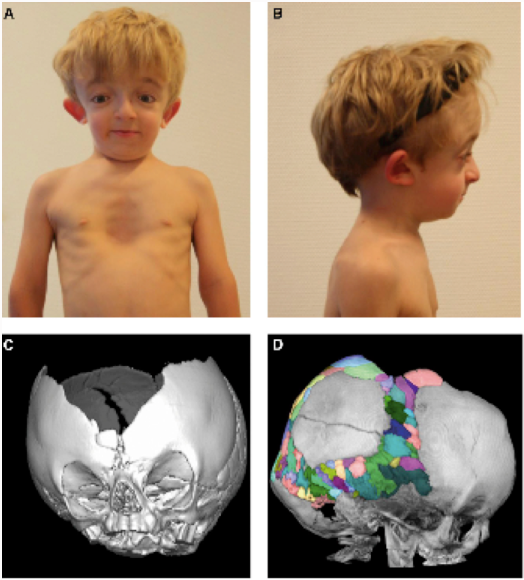
Craniosynostosis, ocular proptosis, hydrocephalus, distinctive facial features, and bone phenotype similar to OI type IV with recurrent diaphyseal fractures.
Images
Fig. Photographs and CT Scans of a patiente affected by Cole-Carpenter syndrome type 2. (A) At the age of 7 years. Note facial dysmorphism with down-slanting palpebral fissures, hypertelorism, dysplastic right ear, and pectus excavatus. (B) Lateral view. Note mid-face hypoplasia, micrognathia, frontal bossing, and dysplastic right ear. (C) Three-dimensional CT scan of the cranium at the age of 14 months. Note wide sagittal suture with a broad ossification defect frontoparietal apical. Sutura metopica is displaced to the bottom, as indicated by surrounding wormian bones. Coronal sutures, lambda sutures, and temporal sutures appear narrow and with premature ossification pattern without clear synos- tosis, but with multiple wormian bones enclosed. (D) Lateral view of a cranial CT scan taken at age 4 years and 4 months, with multiple wormian bones highlighted in different colors. Note an intraparietal suture on the left side (described as unilateral intraparietal suture). The large ossification defect fronto-parietal apical persisted. The sphenoid wings appear with an increased density and with multiple erosions.
Reproduced from Am J Hum Genet, Vol 96, Garbes L, Kim K, Riess A, et al. Mutations in SEC24D, encoding a component of the COPII machinery, cause a syndromic form of osteogenesis imperfecta, Pages 432-9, Copyright 2015, with permission from Elsevier.
Cole-Carpenter syndrome is an extremely rare form of bone dysplasia. It was described for the first time in 1987, by Cole and Carpenter, as a new variant of OI (see also OI type I).
(OMIM phenotype number #112240)
Cole-Carpenter syndrome is an extremely rare form of bone dysplasia. It was described for the first time in 1987, by Cole and Carpenter, as a new variant of OI (see also OI type I). In addition to severe bone fragility, the main features of the syndrome are craniosynostosis, communicating hydrocephalus, ocular proptosis, marked postnatal growth failure, and distinctive facial appearance.
Gene
P4HB gene, 17q25.3 (OMIM gene/locus number #176790). P4HB gene encodes protein disulfide isomerase (PDI).
Phenotype
Craniosynostosis, ocular proptosis, hydrocephalus, distinctive facial features, and bone phenotype similar to OI type IV with recurrent diaphyseal fractures.
Images
Fig. Radiographic Findings in Individual 2 at 18 years of age affected by Cole-Carpenter syndrome type 1. (A) Lateral skull radiograph showing severe midface hypoplasia. (B) The right upper extremity is severely deformed. (C) In the lower extremities, both femurs and tibias have undergone intramedullary rodding surgery. Epiphyses of both distal femurs are wide and seem filled with nodular structures (‘‘popcorn epiphyses’’). The right femur shows a large cystic area (asterisk) and no bone is visible in the mid- shaft area (arrow). (D) Wide epiphyses of the metacarpal and digital bones, thin cortices and a cystic appearance. Some of the end phalanges seem to be partially resorbed (arrows).
Reproduced from Am J Hum Genet, Vol 96, Rauch F, Fahiminiya S, Majewski J, et al. Cole-Carpenter syndrome is caused by a heterozygous missense mutation in P4HB, Pages 425-31, Copyright 2015, with permission from Elsevier.
Focal dermal hypoplasia, or Goltz-Gorlin syndrome, is a rare syndrome, which may affect many different organs such as eyes, bones (limb malformation, osteopathia striata, costovertebral abnormalities, slipt sternum, fibrous dysplasia of bone, syndactyly, polydactyly, “lobster-clawlike” oligodactyly, short stature), teeth, and skin.
(OMIM phenotype number #259770)
Osteoporosis-pseudoglioma syndrome, autosomal recessive, (OPPG) is a very rare disease caused by homozygous or compound heterozygous mutation in the gene encoding low density lipoprotein receptor-related protein-5 (LRP5), with loss of LRP5 function and decreased osteoblast activity. OPPG is characterized by congenital or infancy-onset blindness and extremely severe childhood-onset osteoporosis (lumbar spine Z-score often <-4) with spontaneous fractures. Radiographs disclose severe diffuse demineralization and often reveal fractures. The eye phenotype is secondary to defective vascularization and ranges from congenital phthisis bulbi to milder vitreoretinal changes. Ocular and skeletal abnormalities predominate, although other organs may be involved also. Other clinical manifestations may include: microphthalmos, abnormalities of the iris, lens or vitreous, cataracts, short stature, microcephaly, ligamental laxity, mental retardation and hypotonia. Usually mental development is normal but ∼25% have cognitive impairment. The phenotype is variable even among siblings. The prevalence is approximately 1/2.000.000.
Few data are available on the treatment of osteoporosis in OPPG, however some beneficial response to bisphosphonates have been described.
Gene
LRP5 gene, 11q13.1 (OMIM gene/locus number *603506)
Phenotype
Blindness, microphthalmia, vitreoretinal abnormalities, cataract, phthisis bulbi, absent anterior eye chamber, iris atrophy, pseudoglyoma, muscle hypotonia, obesity, mental retardation (in some cases), ligament laxity, severe osteoporosis, multiple fractures, short stature, kyphoscoliosis, hyperextensible joints, and wide metaphyses.
Images
Fig. (a) Persistence of the fibrovascular system (pseudoglioma) visible upon funduscopy. (b) Standard radiograph of the lumbar spine in a 37-year-old man with OPPG. Note the extremely severe demineralization and multiple vertebral fractures (Levasseur R, Lacombe D, de Vernejoul MC. LRP5 mutations in osteoporosis-pseudoglioma syndrome and high-bone-mass disorders. Joint Bone Spine. 2005 May;72(3):207-14.).
Reproduced from Br J Ophthalmol, Wilson G, Moore A, Allgrove J, volume 85, page 1139, copyright notice 2001 with permission from BMJ Publishing Group Ltd
Other resources:


